
Call Now
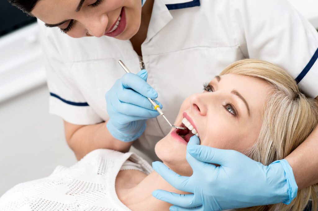
Greenwich Oral Surgery provides comprehensive oral pathology services, combining advanced diagnostic technology with extensive surgical experience. Our oral and maxillofacial surgeons are trained to recognize subtle changes indicating pathological processes, performing thorough evaluations and biopsies when necessary. We work closely with specialized pathology laboratories and oncology teams to ensure accurate diagnosis and appropriate treatment for every patient presenting with oral abnormalities.
Common signs and symptoms to monitor: oral pathology manifests through various warning signs that warrant professional evaluation. Persistent white or red patches inside your mouth, sores that don't heal within two weeks, unexplained lumps or thickening of oral tissues, and chronic pain or numbness in your mouth or jaw all require examination. Changes in how your teeth fit together, difficulty swallowing or moving your tongue, and persistent hoarseness or sore throat may also indicate underlying pathology. Regular self-examination of your mouth, combined with routine dental visits, allows for early detection when treatment is most effective.
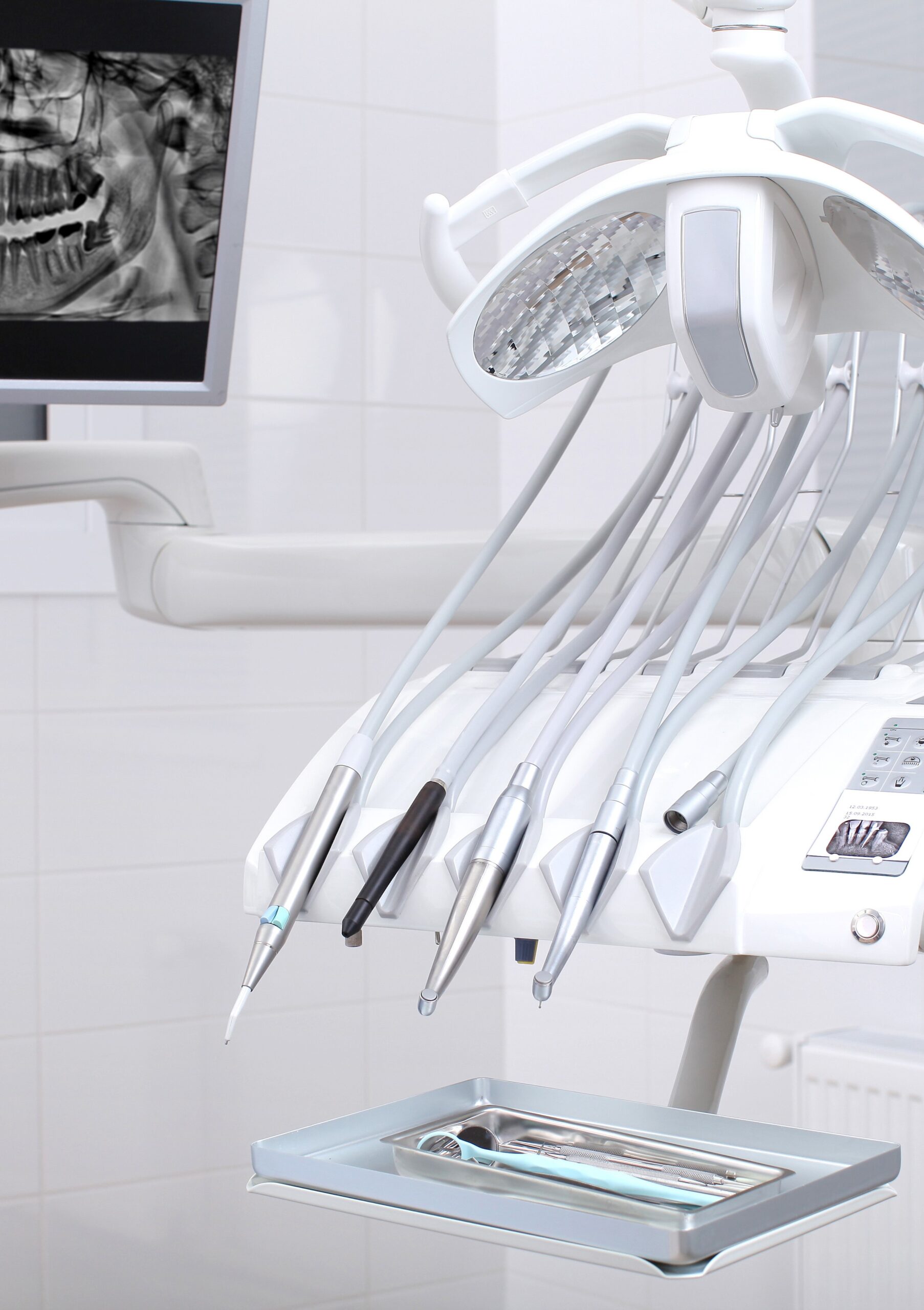
Our diagnostic process begins with a comprehensive clinical examination of all oral tissues, including visual inspection and palpation of suspicious areas. Advanced 3D cone beam CT imaging reveals bone involvement and helps determine lesion extent. When pathology is suspected, we perform biopsy procedures to obtain tissue samples for laboratory analysis. Incisional biopsies remove a portion of abnormal tissue, while excisional biopsies remove the entire lesion. All specimens are sent to specialized oral pathology laboratories for microscopic examination and definitive diagnosis.
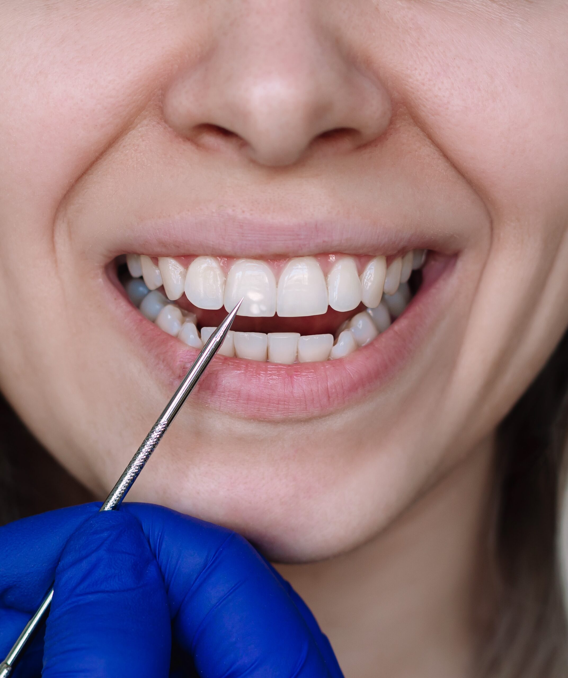
Our surgeons diagnose and treat a wide range of oral pathological conditions. Benign cysts and tumors, including dentigerous cysts and ameloblastomas, require surgical removal and monitoring. Oral mucosal lesions such as leukoplakia and erythroplakia need biopsy evaluation and management. Salivary gland pathology, including blocked ducts and salivary stones, may require surgical intervention. We also manage jaw bone lesions and other abnormal growths requiring excision. When biopsy results indicate serious pathology, we coordinate care with specialists and provide comprehensive treatment planning.
Biopsy procedures allow definitive diagnosis of suspicious oral lesions through microscopic tissue examination. Incisional biopsies remove a small portion of abnormal tissue for analysis, typically performed under local anesthesia in our office. Excisional biopsies completely remove smaller lesions along with a margin of healthy tissue, serving both diagnostic and therapeutic purposes. The procedure takes 15-30 minutes depending on lesion size and location. Tissue samples are immediately sent to specialized oral pathology laboratories for microscopic examination. Results typically return within 7-10 days, at which point we schedule a follow-up consultation to discuss findings and recommend appropriate treatment if needed. Most biopsy sites heal within two weeks with minimal discomfort.
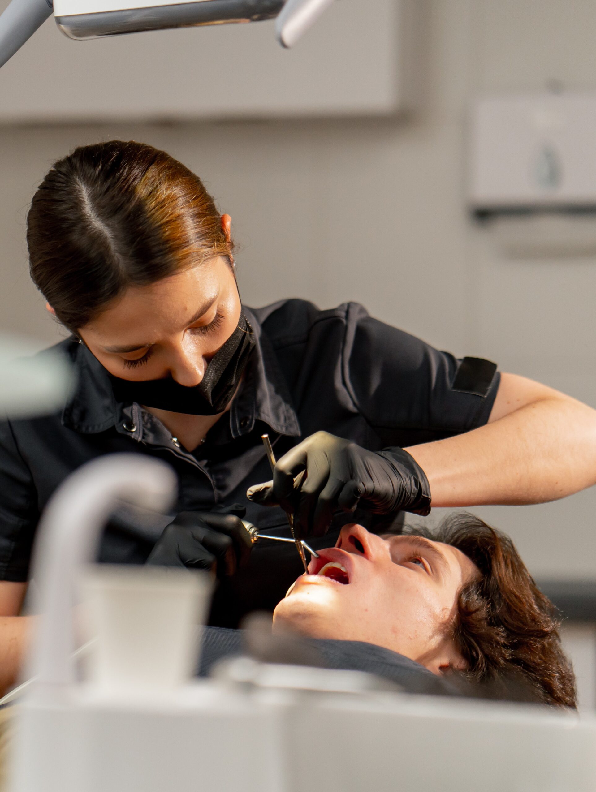
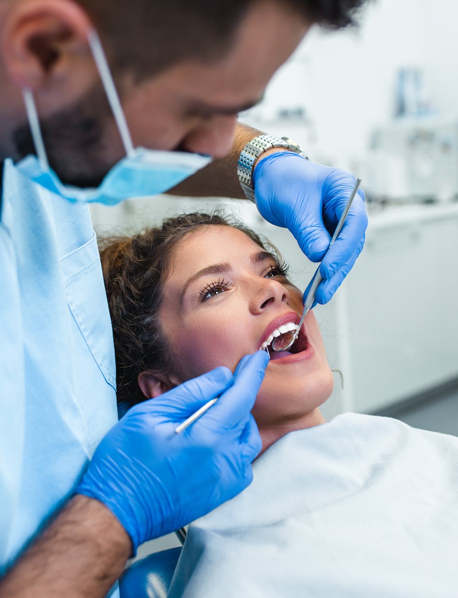
While not all oral pathology is preventable, understanding risk factors helps you reduce your chances of developing serious conditions. Tobacco use in any form—including cigarettes, cigars, and smokeless tobacco— significantly increases risk of oral lesions and malignancies. Heavy alcohol consumption, particularly when combined with tobacco, compounds these risks. Excessive sun exposure contributes to lip pathology. Human papillomavirus (HPV) infection has been linked to certain oral conditions. Maintaining good oral hygiene, attending regular dental examinations, and performing monthly self-examinations of your mouth enable early detection when treatment is most effective. If you notice persistent changes in your oral tissues, seek professional evaluation promptly rather than waiting for symptoms to worsen.